Choose timezone
Your profile timezone:


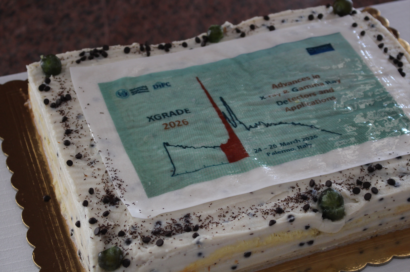
We are pleased to announce the first edition of Advances in X-ray and Gamma RAy DEtectors and applications (XGRADE) workshop that will be held at University of Palermo, Palermo, Italy on 24-26 march 2026.
XGRADE2026 is the first event organized in the framework of the COST Action CA24131-ENRICH: European Network for radiation-detection based Research and Innovation addressing increasing societal CHallenges.
The main goal of XGRADE2026 workshop is to provide a forum for discussion of the state of the art for X-ray and gamma ray detector technology, including materials improvement, device development and characterization, electronic readout, signal processing, system development and applications. The XGRADE workshop will stimulate new ideas and collaborations among researchers working on radiation detectors and imagers for X-ray and gamma ray measurements.
X-ray and gamma ray detectors are finding increasing applications in such diverse fields as medicine, homeland security, radiography, astrophysics, nuclear physics and environmental monitoring.
The venue of the workshop is the Department of Engineering, University of Palermo, Viale delle Scienze, Palermo, Italy.
Main Topics
Authors are invited to submit abstracts presenting their work on one of the topics below:
Conference Chairs
Scientific Advisory Committee
Local Organizing Committe:
High flux CZT detectors show peculiar physical properties that enable several applications in hard X-ray energy range . However, the particular electronic structure of high flux crystals requires a dedicate contact technology. In this work, we describe the technology improvements carried out on the preparation of high quality contacts, as well as several experiments that show the high homogeneity of the material and the high-flux performances. Also, we will show advanced CZT linear array detectors featuring sub-keV energy resolution, fabricated using high-flux HF-CZT material. These devices, specifically optimized for energy-resolved X-ray imaging, achieve a spatial resolution of 500 μm and an energy resolution better than 0.6 keV FWHM at 60 keV, enabling highly accurate spectral discrimination. These activities work were supported by the Italian Ministry for University and Research (MUR), under PRIN 2022 and PRIN 2022-PNRR projects (CUPs: B53D23004720006; B53D23024100001; B53D23004710006; B53D23008550006) and by the ASI contract n. 2023-22-U.0, CUP: C33C23000620005.
The peculiar physical properties of Silicon Carbide (SiC) such as its wide band gap (up to 3.2 eV), high saturation velocities of the charge carriers (200 um/ns), high breakdown field (2 MV/cm), high thermal conductivity (4.9 W/cm²) and large threshold displacement energy (21-35eV), joined with the microelectronics advanced fabrication technology, make this semiconductor unique for X-ray detection and spectroscopy with high energy resolution even above room temperature and in harsh environments. Relevant R&D on SiC X-ray detectors have been made in the last 25 years, and this presentation presents a review of the state of the art reached with different crystals (semi-insulating or epitaxial) and detector types (pad, pixel, microstrip). The present technological limits to be overcome, the challenges and perspectives for further developments will be discussed.
The astrophysics division of CEA is developing high reliability CdTe based pixelated imaging spectrometers with high energy resolution and high dynamic range for astronomy and solar physics in space. The detector technology, known as Caliste, was developed as part of a long-standing research and development program involving detector simulations and performance assessments, the development of custom ASICs and 3D packaging technologies.
This presentation will cover the fundamentals of the Caliste technology and showcase its successful application in space, as demonstrated by STIX (Spectrometric Telescope for X-ray Imaging) aboard the Solar Orbiter ESA mission and MeDDEA (Measuring Directivity to Determine Electron Anisotropy) aboard the PADRE NASA CubeSat, which were launched in 2020 and 2025, respectively. Both instruments use Caliste-SO CdTe-based hard X-ray spectrometers, combining their data to co-observe intense solar flares from different perspectives and two different orbits. This provides unique insights into detector performance in flight.
I will also describe more recent developments towards an ultra-fine pitch CdTe based photon counting imaging spectrometer. This development involves new full-custom front-end ASIC readout and a 3D hybridization concept. The prototype detector assembly on top of our ASICs has been successfully operated and early results will be illustrated. These new developments are paving the way for future scientific space missions.
Some direct societal applications of Caliste technology in the field of nuclear monitoring will be presented as an example of the translation of fundamental research instrumentation to the market.
On the behalf of a wide collaboration.
High energy polarimetry, as recently demonstrated by NASA’s IXPE mission in the soft X ray band, is opening new perspectives in high energy astrophysics by adding a fresh observational dimension alongside the well established techniques of spectroscopy, imaging, and timing. This measurement capability had already been proposed to the scientific community by a broad collaboration in response to the ESA call (1 June 2004) for scientific themes for the preparation of the Cosmic Vision 2015–2025 programme (“X and gamma ray polarisation: the ultimate dimension”).
In the late 1990s, our group began exploring the use of CdTe/CZT spectrometers—at the time an emerging technology in high energy astrophysics (e.g., INTEGRAL/IBIS, Swift/BAT)—to develop detectors capable of exploiting the modulation of Compton scattering expected for linearly polarised photons. Since then, together with national and international collaborators, we have designed several prototypes and proposed various configurations of highly segmented CdTe/CZT spectrometers able to operate as Compton scattering polarimeters for hard X rays and soft gamma rays, while simultaneously providing spectroscopy, timing, and imaging capabilities.
This effort has been accompanied by a series of experiments that have confirmed the suitability of such detectors for polarimetric measurements in the energy range from a few hundred keV up to the MeV domain.
In this presentation, on behalf of all colleagues who have contributed to this long term endeavour, I will outline the development of these activities, summarise the main results obtained over the years, and discuss potential opportunities for future space missions dedicated to hard X ray and soft gamma ray astrophysics.
As transient sky polarimetry gains prominence in high-energy astrophysics, the SWIPE (Soft-X and gamma-ray WIde-field Polarimeter) mission concept introduces an integrated approach to photoelectric and Compton detection. In the 0.2–8 keV range, the mission employs gas mixture photoelectric absorption to maximize sensitivity and efficiency. For higher energies (20–1000 keV), the Compton scattering techniques are usually preferred. The SWIPE Compton polarimeter consists of an array of plastic and inorganic scintillator bars equipped with SiPMs or SDDs at both bar ends. While conventional Compton polarimeters typically reconstruct events using only the azimuthal scattering angle—thereby limiting off-axis performance—SWIPE’s readout configuration allows for Depth of Interaction (DOI) estimation. By analyzing the intensity ratio of the dual signals, the polar scattering enhancing sensitivity and mitigating systematic error. Here we present the SWIPE Compton polarimetry configuration alongside ongoing experimental activities dedicated to prototype development.
Radiotherapy is one of the pillars of cancer treatment, relying on the precise delivery of ionizing radiation to destroy tumor cells while preserving healthy tissue. Central to this balance is dosimetry, which directly influences both therapeutic efficacy and patient safety. As advanced techniques such as VMAT and emerging modalities like FLASH and spatially fractionated radiotherapy push the boundaries of dose delivery, the demand for innovative, high-performance sensors has never been greater. This lecture highlights recent advancements in dosimetry, focusing on the development and application of next-generation detectors. We will explore the evolving roles of new ion chambers, scintillating dosimeters, and solid-state sensors in meeting the challenges posed by modern treatment modalities.
Positron Emission Tomography (PET) imaging is based on measurement of energy and coincidence time of back-to-back gamma photons from positron annihilation. The annihilation quanta are entangled, and this fact is reflected in orthogonality of their initial polarizations, however it has not been utilized in clinical systems. We developed a demonstrator setup based on Compton polarimeters to measure the polarization correlations of annihilation quanta, and to assess the potential of this additional information to enhance PET. Two detector modules, comprising GAGG scintillator matrices with 3.2 mm pitch read out by silicon photomultipliers were used, and they performed with an energy resolution < 9.5% at 511 keV and coincidence time resolution of about 300 ps (FWHM). PET imaging tests done with source of clinically relevant activities from 90 MBq to 400 MBq, show that a spatial resolution from 3.6±0.3 to 4.9±0.3 mm can be achieved in the central field of view, using the polarization-correlated Compton events. We show that these events exhibit up to 20% higher signal to random background ratio, compared to conventional single-pixel events. Finally, we discuss how this concept can be advanced towards clinical application.
An HPGe detector equipped with a transistor reset preamplifier and readout with a CAEN DT5781 fast pulse digitizer was employed in the measurement of X-rays from kaonic lead at the DA𝛷NE 𝑒+𝑒− collider at the Laboratori Nazionali di Frascati of INFN.
We will present results which show that the resolution of the HPGe detector in regular beam conditions remains the same as that without the beam and that a satisfactory background reduction can be achieved.
This feasibility measurement serves as a test bed for future dedicated measurements of kaonic X-rays with an HPGe detector from heavy targets.
The Jagiellonian Positron Emission Tomograph (J-PET) represents the first modular and portable PET scanner capable of multi-photon detection. This multidisciplinary system serves dual purposes in medical imaging and fundamental physics research, including investigations of discrete symmetry violations in positronium decay processes [1,2,3]. Beyond conventional PET imaging capabilities, the J-PET enables positronium lifetime imaging within the human body [4,5,6]. The recent achievement of capturing the first in-vivo positronium image in human brain tissue using J-PET technology [6] demonstrates the promising future of this novel diagnostic approach. The most suitable radiopharmaceutical for this type of imaging would be one with the 44Sc radioisotope [7], and work is currently underway on the preparation of a Scandium generator [8] and the development of this tracer.
My presentation will cover research conducted with the J-PET facility spanning both fundamental physics studies and medical imaging applications.
[1] P. Moskal et al., Nature Communications 12, 5658 (2021).
[2] P. Moskal et al., Nature Communications 15, 79 (2024).
[3] N. Chug et al., Phys. Rev. D 113, 032003 (2026).
[4] P. Moskal et al., Nature Reviews Physics 1, 527 (2019).
[5] P. Moskal et al., Science Advances 7, eabh4394 (2021).
[6] P. Moskal et al., Science Advances 10, eadp2890 (2024).
[7] M. Das et al., IEEE Transactions on Radiation and Plasma Medical Sciences 2025.
[8] P. Moskal et al., Acta Phys. Polon A 148 (6), 152 (2025).
The PANDORA (Plasmas for Astrophysics, Nuclear Decay Observation and Radiation for Archaeometry) project aims to establish a novel experimental facility at INFN-LNS (Catania) dedicated to interdisciplinary research on magnetically confined plasmas, with applications ranging from nuclear astrophysics to radiation sensor and detector testing.
The facility is designed to reproduce selected stellar thermodynamic conditions, particularly in terms of plasma temperature. A primary scientific objective is the first direct measurement of nuclear β-decay rates in hot plasmas. Both theoretical studies and experiments on fully stripped ions have demonstrated that β-decay lifetimes may vary by orders of magnitude in highly ionized environments. The PANDORA setup is based on a compact superconducting B-minimum magnetic trap, where plasmas of different elements are generated via Electron Cyclotron Resonance (ECR). The plasma parameters are expected to reach electron densities nₑ ≈ 10¹¹–10¹³ cm⁻³ and tunable electron temperatures Tₑ ≈ 0.1–30 keV. Under these conditions, the plasma emits intense RF, visible, UV, X-ray, and γ radiation. To fully characterize the plasma state and correlate it with nuclear decay measurements, PANDORA integrates a comprehensive multi-diagnostic system, including RF interferometry and polarimetry, optical and X-ray spectroscopy, X-ray imaging, and spatially resolved spectroscopy performed with different detector technologies. An array of 14 HPGe detectors will be employed to measure β-decay rates through high-resolution γ-ray spectroscopy, detecting the de-excitation photons emitted by the daughter nuclei. The decay rate will be monitored in real time and correlated with the simultaneously measured thermodynamic plasma parameters. The facility is currently under construction, with first plasma expected in late 2026. Beyond β-decay studies, the setup will enable additional astrophysically relevant experiments, such as laboratory measurements of optical opacities under conditions pertinent to r-process nucleosynthesis in kilonova scenarios. Among more than one hundred identified physics cases, the first experimental campaign will focus on a shortlist including ⁹⁴Nb (t₁/₂ ≈ 2 × 10⁴ y), 134Cs (t₁/₂ ≈ 2.5 y), and 176Lu (t₁/₂ ≈ 3.76 × 10¹⁰ y). The facility will also serve as an open platform for testing innovative technologies and methodologies relevant to plasma-based research and will be accessible to the broader scientific community.
Abstract:
Introduction: Intraoperative gamma probes are essential for localizing radiolabeled tumors and sentinel lymph nodes. However, current fixed-geometry designs suffer from a static trade-off between sensitivity and spatial resolution: high sensitivity is required for rapid lesion detection, while high spatial resolution is critical for precise margin delineation. This work presents the design and validation of a novel, reconfigurable LaBr₃(Ce)-SiPM gamma probe capable of intraoperatively switching between these two performance modes.
Methods: We developed a dual-layer lead collimator assembly defined by six geometric parameters. A validated analytical model, coupled with a multi-objective genetic algorithm (NSGA-II), was employed to explore the theoretical performance limits and identify optimal geometries. The framework was validated against MCNP6 simulations and experimental measurements from 123 distinct physical collimator configurations using a ⁹⁹ᵐTc source. A two-phase computational search was conducted to identify a single "universal" geometry that maximizes tunability.
Results: The optimization identified a universal design capable of adapting to clinical requirements via mechanical adjustment of the collimator layer positions. In High-Sensitivity (HS) mode, the probe achieves a sensitivity of 1483 cps/MBq (at 30 mm SCD), facilitating rapid initial searching. In High-Resolution (HR) mode, the system achieves a spatial resolution of 6.41 mm FWHM (at 30 mm SCD) with adequate sensitivity (206 cps/MBq), enabling precise separation of closely spaced nodes and tumor margins. The LaBr₃(Ce) detector provides superior energy resolution (<7% at 140.5 keV), allowing for effective discrimination between ⁹⁹ᵐTc and ¹²³I.
Conclusion: We successfully demonstrated a single-instrument solution that overcomes the limitations of fixed-collimator probes. This reconfigurable architecture offers surgeons the flexibility to prioritize either detection speed or localization precision on demand. This work has been published in Physics in Medicine & Biology (2026), DOI: 10.1088/1361-6560/ae387e.
High purity germanium (HPGe) detectors remain the state of the art technology for high resolution gamma ray spectroscopy, yet conventional lithium diffused contacts impose intrinsic limitations on device performance and segmentation due to their ~1 mm inactive layer and poor thermal stability. To address these constraints, a new fabrication approach based on magnetron sputtered dopants incorporated into germanium via Pulsed Laser Melting (PLM) has been developed at INFN-LNL and the University of Padova. PLM temporarily melts a thin Ge layer, enabling epitaxial regrowth and substitutional dopant incorporation, resulting in an ultra thin, thermally stable, and fully segmentable contact/junction.
A series of prototypes from 2 mm devices to 2 cm thick crystals was fabricated to validate the technique. Across all samples, PLM formed junctions exhibited excellent thermal stability, including after neutron damage recovery annealing cycles. Process optimization (surface preparation, photolithography, chemical mechanical polishing, and mechanical contacting) yielded devices with breakdown voltages significantly above depletion voltage.
These results demonstrate that PLM based doping can effectively replace lithium diffused contacts, enabling next generation segmented HPGe detectors with improved performance for applications in nuclear physics, imaging, security, and astrophysics.
Segmented high-purity germanium (HPGe) detectors are essential tools in advanced gamma spectroscopy. However, many of their technical and manufacturing details remain proprietary to industry. To overcome this limitation, the Italian Institute for Nuclear Physics funded the N3G Project, aimed at developing new segmented HPGe detectors while building in-house expertise for their design, production, operation and maintenance.
In this framework, we present the design and qualification of a cryogenic capsule conceived to host a coaxial high-purity germanium detector and to operate it under high-vacuum conditions at liquid nitrogen temperature. Particular attention is given to mechanical robustness, thermal compatibility and long-term vacuum integrity.
A comprehensive qualification workflow is described, including helium mass spectrometry leak detection, local helium sniffing tests and iterative optimization of the sealing through controlled fastening adjustments. Particular emphasis is placed on the sealing strategy, based on metal gaskets such as HelicoFlex seals, selected for their robustness and suitability in high-vacuum and cryogenic environments. The role of non-evaporable getters is also discussed, highlighting their function as chemical pumps capable of enhancing and stabilizing the static vacuum level over extended operation periods.
The sealing tests demonstrated excellent leak-tight performance: over a five-day monitoring period, the internal pressure increased by only about 2e−5 mbar, confirming the high integrity of the adopted sealing solutions. These results validate the effectiveness of the combined approach—metallic seals and getter-based vacuum stabilization—and provide a solid and reproducible technological basis for future detector developments.
Finally, the mechanical support structures of the detector are described in detail. These components have been specifically designed to ensure stable and safe handling of the device, while minimizing mechanical stress and avoiding any direct contact that could damage its sensitive surfaces. The support system enables the detector to be oriented in any spatial configuration, providing full flexibility during installation, testing and operation, without compromising its structural integrity or performance.
KETEK is the world market leader for Silicon drift detectors (SDDs). Among all market competitors KETEK offers by far the largest product variety associated with SDD modules, operating electronics and detector accessories.
KETEK’s SDDs represent the state of the art in energy dispersive X-ray fluorescence detection in the energy range from about 0.05 keV to about 30 keV. They are used worldwide in a large variety of applications, including electron microscopes, benchtop and handheld XRF spectrometers. Thanks to their wide operating temperature range, excellent energy resolution, and high reliability, they are well suited for both industrial applications and research.
This presentation provides an overview of KETEK’s holistic system approach, covering everything from the innovative graphene entrance window and specialized detector concepts to readout electronics and software. Featured developments include stray-line-free SDDs for improved spectral purity, rise-time-optimized detectors with large active areas, digital signal processing with very short peaking times for high throughput applications as well as multi-channel detector systems with a total collimated detection area up to 350 mm². This all is complemented by stable and user-friendly software. KETEK’s solutions range from fully integrated systems in compact, robust housings to miniaturized, modular systems optimized for integration into customer-specific measurement setups.
The VOXES (VOn Hamos X-ray spectrometer using HAPG for Extended Sources) Bragg spectrometer is a high-resolution X-ray spectrometer developed at the INFN National Laboratories of Frascati (LNF). It is based on cylindrically bent mosaic crystals operating in the Von Hamos configuration and was specifically designed to overcome the intrinsic efficiency limitations of traditional Bragg spectroscopy when dealing with extended and diffuse X-ray sources. By using Highly Annealed Pyrolytic Graphite (HAPG) crystals with typical mosaicity of 0.007–0.01°, optimized beam optics, and a compact geometry (source–crystal distance ~50–100 cm, crystal radius ~100–200 mm), VOXES achieves energy resolutions of about 2–5 eV (σ) in the 5–10 keV range, corresponding to resolving powers E/ΔE up to ~2000–3000. Absolute energy precision at the level of ~0.1–0.2 eV can be obtained after calibration, while maintaining high collection efficiency even for effective source sizes of a few mm in the dispersive plane, and up to a few cm in the vertical one. The spectrometer provides simultaneous multi-line detection over energy windows of ~100–2000 eV, depending on geometry and detector, and demonstrates detection efficiencies orders of magnitude higher than traditional perfect-crystal Bragg systems when measuring extended or low-intensity sources.
Since its first prototype, VOXES has undergone continuous development, including improvements in ray-tracing-based optimization, efficiency characterization, and calibration procedures. Recent upgrades introduced enhanced automation, motorized positioning with micrometric precision, and the integration of an energy-dispersive fluorescence monitoring line using a silicon PIN-diode detector, enabling simultaneous wavelength- and energy-dispersive measurements and real-time flux normalization. Dedicated sample environments, including liquid-sample holders and transmission geometries, allow laboratory X-ray emission and (in future) absorption studies with count rates ranging from a few Hz up to several kHz, depending on configuration and source intensity.
VOXES has been successfully employed in multiple research programs. In fundamental and nuclear physics, it has been developed as a precision instrument for high-resolution spectroscopy of exotic-atom X-ray transitions and related measurements. Within the TASTE (INFN-CNTT) and MITIQO project (Regione Lazio), VOXES demonstrated high-resolution X-ray emission spectroscopy of transition metals in liquid solutions, enabling determination of oxidation states and chemical environments with energy sensitivities of a few tenths of an eV.
Today, VOXES integrates multiple detectors, including MYTHEN2 (50 µm pitch strip detector), Timepix3 (55 µm pixel, event-driven readout with 1.56 ns time resolution), a CCD detector, and a silicon PIN diode, allowing flexible spectroscopic 1D and 2D measurements. A large collection of HAPG and Silicon crystals enables coverage of a wide photon-energy range from approximately 4 to 40 keV. With its modular design, high resolving power, and ability to operate with extended and weak sources, VOXES represents a versatile laboratory-scale spectroscopic facility bridging fundamental physics, materials science, and applied X-ray research.
In the frame of the PANDORA project - aimed at measuring β-decays of nuclear astrophysical interest in magnetically confined plasmas – and the SAMOTHRACE ecosystem (funded by the EU Next Gen Program), a new non-invasive plasma diagnostics testbench, PYN-HO, has been developed at INFN-LNS to advance the state-of-the-art in non-invasive plasma monitoring. The system is designed for high-resolution X-ray imaging, space-resolved spectroscopy, and tomography, in the 0.4–30 keV range.
The setup features a 4 MP X-ray CCD camera coupled with a 400 µm Pb pinhole, a multi-collimation system for scattering suppression, and a millisecond-resolution Pt-Ir X-ray shutter. Advanced Single Photon-Counting (SPhC) and High-Dynamic-Range (HDR) algorithms are employed to perform space-resolved spectroscopy and spectrally-resolved imaging, enhancing image quality and removing readout noise.
The system's capabilities were recently demonstrated in experiments at the ATOMKI laboratory (Debrecen, Hungary), where plasma transients—including ignition, afterglow, and turbulence—were resolved with 460 µm spatial, 230 eV (at 8 keV) energy, and 100 µs temporal resolution through dedicated trigger systems and delays. A SDD was simultaneous used to benchmark the spectroscopic results. These measurements enabled the characterization of structural and temporal plasma evolution and the investigation of electron/ion deconfinement dynamics between different configurations.
Furthermore, a new algorithm based on Artificial Intelligence and Machine Learning approach is currently being developed for SPhC image analysis, aimed at significantly improving the system’s overall performance and computational speed.
The PYN-HO prototype can be integrated into different facilities to study plasma structure, confinement dynamics, instabilities, in-plasma and plasma vessel elemental composition, and local thermodynamic parameters. These results highlight the potential of advanced X-ray diagnostics in optimizing ion source performance and exploring new frontiers in nuclear astrophysics but are potentially ready also for novel future applications.
Hybrid semiconductor single photon counting detectors developed within the Medipix collaboration at CERN represent a modern detector technology based on the principle of single-particle counting (alpha, gamma, electrons, etc.). A characteristic feature of these detectors is that each pixel contains its own readout electronics (amplifier, discriminator, etc.), which eliminates the integration of additive noise typical of conventional integrating detectors (e.g., flat panels, CMOS). These detectors consist of two separate parts – a sensor chip (Si, CdTe, etc.) and a readout chip (ASIC), which are interconnected using bump bonding technology. Depending on the generation of the readout chip (ROC), the detector provides comprehensive information including the energy of the interacting particle, the time of its interaction with the detector, and the precise spatial localization of this interaction.The readout chip is composed of a 256 × 256 pixel matrix with a pixel size of 55 µm and can be combined with various sensor materials of different thicknesses depending on the specific application.
The main advantages of these detectors include high spatial and temporal resolution, the ability to perform energy discrimination, virtually unlimited dynamic range, minimal noise, and the possibility of achieving subpixel resolution.
These detectors find wide application both in imaging methods (2D and 3D radiography) and in particle tracking methods, where they enable identification of individual particle types and provide comprehensive information about the radiation field in which the detector is located.
This contribution presents the possibilities of using single-photon counting detector technology and the achieved results across a wide spectrum of applications, such as material analysis using X-ray diffraction, non-destructive testing including applications in nuclear energy, monitoring of radiotherapeutic beams, and soft tissue imaging.
Detector systems utilising the small pixel effect, such as HEXITEC, display high rates of charge sharing due to the similar scales of the charge carrier clouds generated by hard X-ray interactions and the pixel pitch of the detector.
Charge sharing distributes the complete interaction energy of event across multiple readout channels – with losses occurring due to truncation of fractional pixel signals by the energy threshold and inter-pixel trapping. As charge sharing may occur due to charge carrier cloud diffusion/repulsion or multiple photon interaction processes (i.e. detector fluorescence), charge sharing signals degrade the energy and spatial resolution of a detector system.
To preserve system performance, charge sharing signals are typically excluded using Charge Sharing Discrimination (CSD), or the fractional pixel signals are summed without energy-loss or position correction, known as Charge Sharing Addition (CSA) [1]. For photon-limited applications such as medical imaging, CSD results in sensitivity losses of ≈ 67% at 141 keV, whilst CSA significantly degrades energy resolution. The development of charge sharing reconstruction approaches based on the physical process responsible for a charge sharing event allows correction for inter-pixel signal losses and determination of the event interaction position – avoiding the limitations of both CSD and CSA.
Charge sharing interaction process has been demonstrated to correlate with event shape for 2-pixel events [2]. We present a range of energy-analysis methods, based on event shape analysis, which identify the most-likely interaction process responsible for charge sharing events up to 4 pixels in size (characterising >99.996% of events at 141 keV). This allows pre-established methods for energy [3, 4] and position [5] correction to be applied only to events, or sub-processes within complex events, which require correction. Position correction methods for 2-pixel events have also been generalised to allow correction of fractional sub-processes within events. This correction toolkit is sufficient for energy and position correction of all 2-pixel events and most 3-pixel events; achieving improved spatial resolution, 2-3x increased sensitivity when compared with CSD and reduced energy resolution degradation when compared to CSA.
Micro-pattern gaseous detectors (MPGDs) have demonstrated outstanding performance in X-ray and gamma-ray detection; however, further improvements in gain stability, signal formation, and spatial resolution are still strongly connected to the microscopic transport properties of charge carriers in the gas. In this contribution, we present recent developments on nano-patterned GEM (nano-GEM) detectors, focusing on the role of ionic-cluster formation and transport in gas-filled radiation detectors.
The nano-GEM concept exploits nano-structured dielectric and electrode geometries to modify local electric fields and charge transport mechanisms. By combining experimental measurements with detailed simulations using Garfield++ and Magboltz, we investigate how ion clustering phenomena affect signal formation, ion mobility, and space-charge effects under X-ray irradiation. Particular attention is devoted to the impact of nano-scale patterning on detector response, gain uniformity, and temporal characteristics.
Our results indicate that nano-structured GEM architectures provide an effective pathway to control ion transport and mitigate ion-related performance limitations, opening new perspectives for high-rate, high-stability X-ray and gamma-ray detection systems. These findings are directly relevant for next-generation radiation detectors and imaging applications within the framework of the ENRICH COST Action.
Abstract: Cosmic-ray muography has emerged as a valuable technique for the non-invasive inspection of geological and industrial structures, such as the assessment of nuclear reactor fuel. Despite its successes, conventional muon imaging methods have faced significant challenges in resolving small-scale structures composed of low atomic number and low-density materials. To address this limitation, our research group has pioneered a novel imaging approach that utilizes cosmic-ray muon induced secondary radiation produced within the target material. By using secondary particles—detected in coincidence with cosmic-ray muons by plastic scintillator detectors and a dedicated muon tracker—we have achieved the first cosmic-ray muon radiographs of bone and soft tissue. These images represent the first radiographic visualisations of organic structures obtained via cosmic rays.
Our research further employs Monte Carlo simulations, conducted with the Geant4 software package, to model the interactions of cosmic-ray muons with various detectors and target materials. These simulations are essential in optimising experimental configurations and deepening our understanding of the underlying imaging processes.
In this work, we present two experimental setups: the MUCA (Muon Camera) system in Novi Sad and the COMIS (Cosmic Muon Induced Secondaries) system in Budapest. The MUCA setup comprises four plastic scintillation detectors (50 cm × 50 cm × 5 cm) and a muon tracker (five CCC boards, 25 cm × 25 cm) positioned above the object under investigation. The COMIS setup features a muon tracker (five CCC boards, 50 cm × 50 cm, with 2 mm resolution) located beneath the object, four large plastic scintillation detectors (50cm × 50cm × 5cm) arranged around the target, and four plastic scintillation detectors (25cm × 25cm × 5cm) under the target-volume. The aim of this research is to advance imaging and compositional analysis of diverse objects—particularly those with low atomic number and density—using only naturally occurring cosmic radiation.
Keywords: cosmic muon imaging; muon secondaries detection; low atomic number materials; Geant4 simulations; coincidence event detection
Like many developing countries, Libya is recently facing associated environmental and public health challenges related to radiological safety in the natural resources and built environment.
The growing reliance on imported building materials, combined with aging infrastructure, limited ventilation practices, groundwater rising levels, and increasing concerns about water contamination, necessitates a thorough evaluation of radiation exposure pathways. The naturally occurring radioactive materials (NORM) In particular in the construction stones, indoor radon accumulation, and potential radiological pollution of groundwater, represent emerging but it is still under-addressed hazard in the Libyan status.
This contribution combines two interrelated studies. The first estimates the NORM concentration in imported granite and marble building materials used in Libya, evaluating activity concentrations of Ra-226, Ra-228, and K-40, radiological hazard indices, and radon exhalation potential using HPGe system. The second study inspects indoor radon gas distribution in closed and aged offices, schools, and universities in Tripoli, with highlighting the effects of building age, ventilation, occupancy, and materials of construction.
In addition, the study focuses on broader Libyan environmental concerns related to radiation in water resources, including potential groundwater contamination and the lack of regular radiological monitoring of drinking and irrigation water.
Joining XGRADE2026 is not only an opportunity to share these results at an international forum of scientists but also a chance to get familiar with the state of the art radiation detection, monitoring methods, and environmental assessment tools of today. This kind of interaction is necessary for the improvement of Libya's research capacity, the facilitation of technology transfer, and the building of international cooperation. The mentioned results will be used for the purpose of developing national policies for radiation protection, water and infrastructure management that are sustainable, and environmental policy based on the availability of evidence and in concurrence with the international standards.
Luminescence dating accuracy relies on the precise determination of the total dose rate (Dr). While alpha and beta radiation effects are highly localized, gamma radiation forms a complex, continuous field influenced by the heterogeneous distribution of radionuclides throughout a sediment profile. Standard dosimetry often overlooks these spatial fluctuations, potentially leading to age inaccuracies in stratified deposits.
In this study, we present a spatial modelling approach to map the gamma radiation field within an aeolian sediment profile located near Toruń, Poland. In this early-stage work we propose a model that illustrates how different-activity layers influence more distant sampling points, by accounting for the discrete distribution of natural decay chain radionuclides across the profile, our model.
Simultaneous measurement of interaction position and energy is essential for semiconductor gamma imagers, but optimizing detection efficiency, energy resolution, and spatial resolution presents a significant challenge due to competing trade-offs among noise, signal induction, and readout electronics. While techniques such as cathode readout, pulse shape analysis, and multi-anode readout have been employed for pixelated CdZnTe detectors, digitized signal processing is increasingly leveraged to enhance performance.
To achieve a tileable and modular system, we adopt an anode-only readout scheme combined with multi-anode signal acquisition. Through detailed simulations and digital signal analysis, we investigate the physical factors governing performance, validating our approach with consistent experimental results. This enables a systematic study of signal processing optimization while assessing the impact of key parameters (including readout architecture, detector geometry, bias voltage, and material properties) on spatial and energy resolution.
Finally, we correlate these detector-level optimizations with imaging performance in a high-resolution coded-aperture system, discussing the relative contributions of detection efficiency, spectral and spatial resolution to overall image quality.
We present a novel digital acquisition and processing architecture for high-count-rate X-ray spectroscopy based on the direct digitization of transistor-reset preamplifier outputs. The proposed approach overcomes the limitations of conventional analog reshaping techniques, which suffer from baseline distortions and pile-up effects at high event rates, by employing a high-resolution 20-bit successive approximation register (SAR) ADC with a 6 V input range and a least significant bit of approximately 5 µV. This enables the accurate measurement of millivolt-scale signal steps superimposed on the large voltage ramp characteristic of reset-type charge-sensitive preamplifiers, achieving an effective dynamic range exceeding 1:1,000,000.
To reach the sampling rates required for fast signal processing, four identical 20-bit ADCs operating at 40 Msps are combined in an interleaved configuration, providing an effective sampling rate of 160 Msps. Precise phase alignment, gain matching, and offset correction are achieved through a mixed analog-digital calibration scheme managed by a Zynq System-on-Chip, which also implements real-time digital trapezoidal filtering, baseline estimation, trigger generation, and spectrum building. A dedicated calibration procedure based on a multi-phase sinusoidal injection and Levenberg–Marquardt fitting is used to compensate interleaving mismatches at 20-bit resolution.
The system has been prototyped on a custom acquisition board and experimentally validated using the ARDESIA multipixel silicon drift detector. Preliminary measurements obtained with a single ADC channel demonstrate an energy resolution of 128 eV FWHM at 2 µs peaking time and 140 eV FWHM at an event rate of 1 Mcps with fast shaping. These results confirm the effectiveness of the proposed direct-sampling architecture and demonstrate its suitability for high-rate, high-resolution X-ray spectroscopy in advanced detector systems.
Segmented high-purity germanium (HPGe) detectors are essential tools in advanced gamma spectroscopy. However, many of their technical and manufacturing details remain proprietary to industry. To overcome this limitation, the Italian Institute for Nuclear Physics funded the N3G Project, aimed at developing new segmented HPGe detectors while building in-house expertise for their design, production, operation and maintenance.
This work presents two key outcomes of the project. The first is the development and validation of an innovative electrical interconnection system between the detector electrodes and the front-end electronics, designed to ensure minimal impact on detector leakage current. The solution relies on flexible PCBs fabricated on a 36 $\mu m$ polyimide substrate and shaped to conform to the detector geometry. Its performance was benchmarked against a conventional micro-spring contact scheme with an indium interlayer, demonstrating a reduction in leakage current of more than four orders of magnitude.
The second outcome is a newly designed integrated charge-sensitive preamplifier and its experimental validation. The device is based on a custom Application-Specific Integrated Circuit (ASIC) developed for HPGe signal read-out and implemented in the AMS C35B4C3 technology, a mature and reliable 350 nm CMOS process from Austria Microsystems. Experimental characterization demonstrated excellent performance, including strong linearity (integral non-linearity below 0.03 %) and low noise, with an equivalent noise charge of 108 RMS electrons for a 1 MeV equivalent event and 15 pF electrode capacitance.
Fricke gel (FG) dosimeters have attracted considerable interest in radiotherapy thanks to their capability to map three dimensional dose distributions (DDs). These systems exploit the radio induced oxidation of ferrous (Fe²⁺) to ferric (Fe³⁺) ions, using the concentration of Fe³⁺—detectable via magnetic resonance due to its paramagnetic properties—as a quantitative marker of absorbed dose. Alternatively, by adding a chelating agent such as xylenol orange (XO) or methylthymol blue (MTB), DDs can be retrieved through optical transmittance measurements.
Despite their good sensitivity, linearity, and spatial resolution, the clinical use of FG dosimeters remains limited by their short usable lifetime. Although the gel matrix restricts ion mobility, diffusion still occurs, producing blurring in the measured DD. Moreover, chelating agents induce auto oxidation (AO) processes that alter the relationship between absorbed dose and Fe³⁺ concentration. Both diffusion and AO depend on the time elapsed between preparation/irradiation and measurement, creating a mismatch between FG dosimetry and the logistical constraints of radiotherapy workflows.
Most research in this field has focused on optimizing chemical formulations to mitigate ion diffusion and AO. In this work, we present an AI based post processing technique that enables accurate DD reconstruction when diffusion and AO have already occurred. The method relies on Physics Informed Neural Networks (PINNs): by modelling the underlying physical processes, the network effectively performs a synthetic inversion in time, recovering the original DD from degraded measurements.
We demonstrate that this approach is robust and general with respect to gel composition and problem dimensionality. It yields DDs with low mean squared errors (~10⁻⁶–10⁻⁵ OD²) and high gamma analysis passing rates (>95%). Finally, we show that the effective utilization time of FG dosimeters can be extended by at least an order of magnitude.
The X-ray secondary sources driven by high-power lasers comprise unique features of micrometric source size and femtosecond pulse duration and are of high interest for time-resolved high-resolution X-ray imaging in a wide area of applications of physics, material science and biochemistry [1, 2]. The gas target requirements for optimal laser wakefield acceleration (LWFA) of electrons and efficient secondary radiation are different and depend on available laser pulse energy, pulse duration, requirements of energy and charge of accelerated particles. The formation of suitable gas density profiles in the path of propagation of laser beam are formed by an array of supersonic nozzles producing shaped targets of required plasma concentration. The gas flow of the nozzles is simulated using ANSYS Fluent software. The LWFA of electrons and radiation are simulated using FBPIC Particle-in-Cell and Synchrad code. The nozzles are manufactured using hybrid nanosecond rear-side processing and Femtosecond Laser-assisted Selective Etching (FLSE) machining technologies from single fused silica block [3,4]. The targets were tested experimentally in co-operation with Lund, ELI-Beams, ELI-ALPS and ENSTA LOA laser centres.
The Imaging X-ray Polarimetry Explorer (IXPE) provides unique comprehension about polarized x-ray emissions from many astrophysical sources such as radio quiet and radio loud AGNs,galactic black hole binaries and neutron stars. Polarization maps in X-rays of pulsar wind nebulae and supernova remnants have been probed. IXPE has demonstrated the scientific potential of the techniques but revealed some limitations such as, due to the deposition of ions onto the dielectric of Gas Electron Multiplier (GEM) leads towards the decrease in the gas gain. Also, it has arelatively narrow energy band coupled with rapidly declining efficiency. Another limitation of IXPE GPDs was the large dead time (1.1ms), which is incompatible with the brightest sources.Literature showed that 1-cm drift with an 800-mbar dimethyl-ether gas mixture provides the best polarimetric sensitivity. Further optimization of these parameters depends on the mirror's effective area and the energy of x ray source, but no major improvement is expected with current detector design.
So, to overcome all these limitations and better performance, we are developing a next generation 3-D photoelectron track polarimeter based on Gridpix detector with multipurpose ASICs from the Medipix collaboration, Timepix3 and TimePix4. Gridpix structure in principle prevents the buildup of charge on the metallic mesh, offering a small diffusion with respect to GEM. Also, with the Timepix3 we can record Time-of-Arrival (ToA) and Time-over-Threshold (ToT) simultaneously in each pixel. They would also solve the issue of dead time for any planned high-throughput optics. In this work Gridpix detector experimentally characterized at the INAF-IAPS
laboratory using a novel gas mixture of argon (Ar) and dimethyl ether (DME) (80:20). Combining the argon with the low diffusion and precise tracking capability of DME improves the detector performance. To extract key parameters such as energy spectra, energy resolution, and calibration
from raw experimental data demonstrate a good detector response. For the validation of experimental data, a detailed Geant4 simulation is performed. We present the tuning of the simulation to achieve agreement with experimental measurements obtained using a GridPix prototype, highlighting the importance of accurately modeling both the detector response and the reconstruction methods. These results demonstrate the strong potential of the GridPix detector with an Ar:DME gas mixture as a promising candidate for future high-precision X-ray polarimetry
missions.
Cadmium Zinc Telluride (CZT) detectors are a powerful platform for radiation detection in the keV energy range, combining room-temperature operation and a compact geometry. Their deployment in accelerator environments opens new experimental opportunities, but also introduces complex and structured background conditions that might limit their sensitivities. In this work, we present a Machine Learning driven strategy for background optimization of CZT detectors operated at the DAΦNE accelerator at the INFN National Laboratories of Frascati. Detailed Geant4 simulations of the full experimental setup are used to generate labeled datasets that encode the physical origin and topology of signal and background events. Supervised learning models are trained on these simulations to learn multidimensional correlations that are not accessible through traditional cut-based analyses. We show that ML-based event classification significantly improves background rejection while maintaining high signal efficiency, effectively enhancing the achievable sensitivity of the detector system. This approach demonstrates how simulation-informed learning can become a key tool in the design and optimization of next-generation semiconductor detectors in accelerator-based experiments.
CadTool is an open source simulation framework for x-ray and gamma-ray solid state detectors. The tool uses the SolidStateDetectors.jl library for the simulation of detector electric and weighting fields and implements a modified charge drift model to take into consideration initial charge cloud size, self-repulsion, charge trapping and thermal diffusion. The estimation of the initial charge cloud is done with the simulation of the electron paths inside a given detector material using GATE physics simulation tool. Considering charge dynamics and charge trapping probability we compute Charge Induction Efficiency maps for a 2x2 pixel area. The induced charge/current signals on the cathode and anode electrodes can also be extracted allowing the user to perform parameterization of the transient signals for new electronics design philosophy (ie:. subpixel localization, depth of interaction, timing requirements). To correlate the simulated detector response to real measurement we use GATE to obtain charge deposits on the detector volume according to tunable source geometries and experimental environment. Electronics readout logic models are also implemented to mimic real data acquisition systems (ie:. electronic noise, shaping time, ADC characteristics). The tool has been validated with real detectors operating <100 keV, pixel and double sided strip, and we are actively updating the physics models for the validation of keV - MeV detectors.
The development of high-performance X-ray and gamma-ray detectors is fundamentally constrained by the need to balance detection efficiency, signal stability, material thickness, and long-term radiation tolerance.[1–3] Despite extensive experimental efforts, the existing literature remains fragmented across material classes and device architectures, limiting the establishment of generalizable design principles. In this work, machine learning is employed as an interpretative framework to integrate the experimental data on radiation-sensitive materials and extract physically meaningful performance trends. A curated dataset was constructed from experimental studies including detection efficiency, luminescence yield, response time, radiation-induced degradation, and device architecture under relevant irradiation conditions. Normalized sensitivity and stability indices were derived to enable cross-study comparison. Dimensionality reduction, feature-importance analysis, and unsupervised clustering were applied to map the detector design space. The analysis reveals that the primary performance trade-off is captured by a dominant principal component explaining approximately 55–60% of the variance, corresponding to the balance between signal yield and material thickness, while a second component (20–25%) reflects radiation stability and operational durability. Organic, inorganic, hybrid, and multilayer detector systems occupy distinct performance regimes. Multilayer and hybrid architectures consistently exhibit 15–25% higher thickness normalized sensitivity at comparable stability levels. Feature-importance analysis identifies effective density, interfacial configuration, and emission efficiency as the dominant controlling parameters. These results demonstrate that radiation detection performance is primarily governed by architecture-enabled optimization rather than material composition alone. The proposed machine-learning-driven framework provides quantitative and physically meaningful design guidance for the development of compact, sensitive, and durable X-ray and gamma-ray detectors for potential applications.
Keywords: X-ray detectors; Gamma-ray detection; Machine learning; Hybrid materials; Detector architecture; Radiation stability
References
[1] B. Hou, Q. Chen, L. Yi, P. Sellin, H.-T. Sun, L.J. Wong, X. Liu, Materials innovation and electrical engineering in X-ray detection, Nat Rev Electr Eng 1 (2024) 639–655. https://doi.org/10.1038/s44287-024-00086-x.
[2] M. Chen, C. Wang, W. Hu, Organic photoelectric materials for X-ray and gamma ray detection: mechanism, material preparation and application, J. Mater. Chem. C 9 (2021) 4709–4729. https://doi.org/10.1039/D1TC00525A.
[3] Z. Lin, S. Lv, Z. Yang, J. Qiu, S. Zhou, Structured Scintillators for Efficient Radiation Detection, Advanced Science 9 (2022) 2102439. https://doi.org/10.1002/advs.202102439.